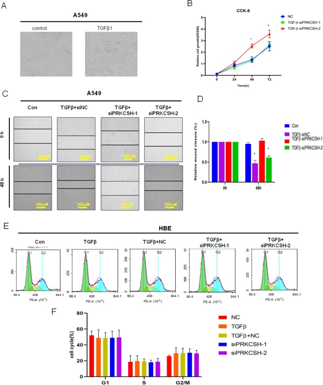
Figure 4.
PRKCSH-2 deficiency promotes lung cancer cell proliferation. A, Representative image of lung A549 cells with or without TGFβ treatment (magnification, ×40). B, Cell Counting Kit-8 (CCK-8) assay was used to determine the effect of silencing the PRKCSH splice variants on the cell proliferation. C, Wound healing assay showed that silence of PRKCSH-2 significantly repressed cell migration ability of A549 cells (magnification, ×10). D, Quantitative determination of wounding healing results. The results represent the average of 3 independent experiments (mean ± standard error of the mean); *P < .05. E, Flow cytometric analysis of cell cycle in 5 groups, control group, TGFβ treatment, TGFβ treatment + negative control (NC), TGFβ treatment + siPRKCSH-1, and TGFβ treatment + siPRKCSH-2. F, Quantitative determination of wounding healing results. The results represent the average of 3 independent experiments (mean ± standard error of the mean); *P < .05.

