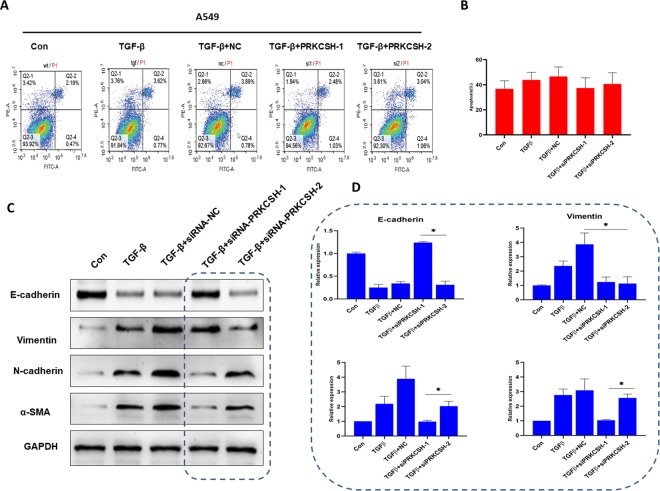
Figure 5.
PRKCSH-2 deficiency promotes TGFβ-induced EMT progression. A, Flow cytometric analysis of apoptosis in 5 groups, control group, TGFβ treatment, TGFβ treatment + negative control (NC), TGFβ treatment + siPRKCSH-1, and TGFβ treatment + siPRKCSH-2. B, Quantitative determination of wounding healing results. The results represent the average of 3 independent experiments (mean ± standard error of the mean); *P < .05. C, Western blotting was used to determine the effects of silence PRKCSH-1 and PRKCSH-2 on the TGFβ-induced EMT progression. D, Quantitative determination of wounding healing results. The results represent the average of 3 independent experiments (mean ± standard error of the mean); * P < .05 EMT indicates epithelial–mesenchymal transition; TGFβ, transforming growth factor beta 1.

