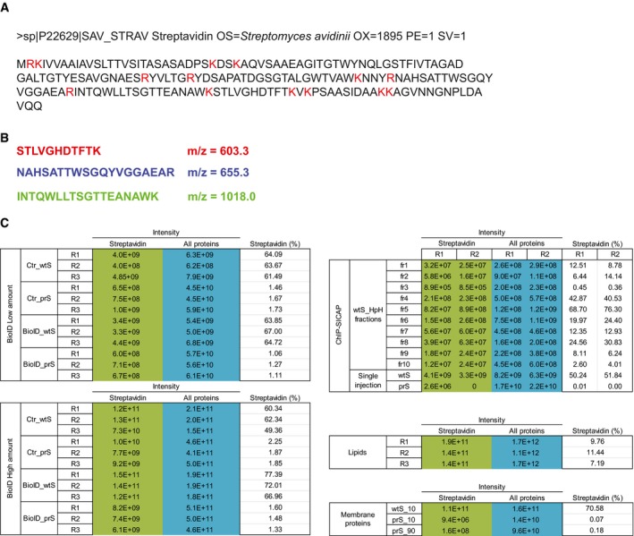
Figure EV1. Tryptic digestion of streptavidin.

- Amino acid sequence of streptavidin protein. Residues cleaved by trypsin are shown in red.
- Sequence and m/z of the top‐3 streptavidin peptides, used to determine the extracted ion chromatogram (XIC) shown in main Fig 1E and F.
