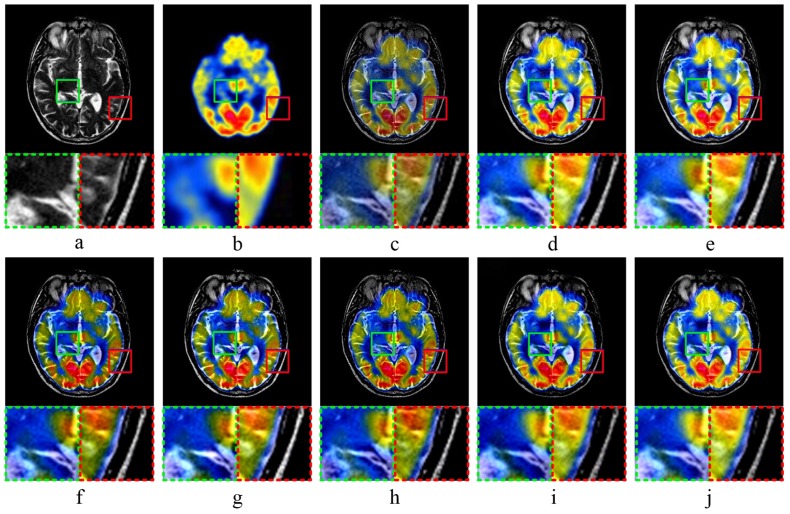
Figure 5.
MR-PET image fusion Experiment 1: (a,b) source images; and (c–j) the fused image obtained by MST-SR, NSCT-PC, NSST-PCNN, ASR, CT, KIM, CNN-LIU, and the proposed method, respectively. Two partially enlarged images marked in green and red dashed frames correspond to the regions surrounded by green and red frames in the fused image.

