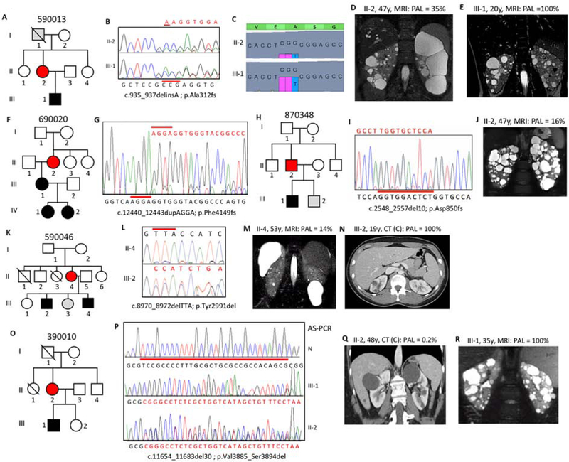
Figure 2. Pedigree, imaging and sequencing data of mosaic families segregating the pathogenic variant to the next generation.
(A) Pedigree 590013 showing ADPKD in II-2 and III-1, with I-1 found to have a few, likely simple, cysts at 79y. (B) SS showing the indel (red line) and reduced peak height of the frameshifted sequence (red) in II-2 compared to III-1. (C) tNGS showing the CCG deletion and T insertion in a reduced number of reads in II-2 compared to III-1. (D) MRI of II-2 at 47y showing asymmetric disease with just a few large cysts in the left kidney compared to a more even distribution in III-1 (E). (F) Pedigree 690020 with ADPKD in three generations. (G) SS of II-2 showing the frameshifted sequence following the 4bp duplication at a low level, reflecting mosaicism. (H) Pedigree of family 870348 with ADPKD in II-2 and III-1. (I) SS of II-2 (reverse strand) showing frameshifted sequence due to a 10bp deletion at a very low level, which is confirmed by AS-PCR (Figure S2B). (J) MRI showing robust PKD in II-2. (K) Pedigree 590046 showing the mosaic case (II-4) with two affected children (III-2 and III-4). (L) The inframe codon deletion is seen at only a low level in II-4 compared to III-2. (M) MRI of II-4 showing very mild kidney and liver cystic disease. (N) Contrast (C) enhanced CT of III-2 at 19y showing a few kidney cysts. (O) Pedigree 390010 shows ADPKD in the II-2 and III-1. (P) SS of the AS-PCR from a normal individual (N), the son (III-1) and the mosaic mother (II-2) shows the 30bp deletion in the son, but due to the low-level mosaicism, the AS-PCR is not completely specific in II-2, hence the doublet sequence. (Q) CT of II-2 shows just a couple of cysts, in contrast to the typical PKD shown by MRI in III-1 (R). Pedigree: red shaded, mosaic; gray, equivocal or unknown; white, ADPKD negative. The percentage of the observed versus the expected level of the pathogenic allele (PAL) is indicated next to each radiological image.

