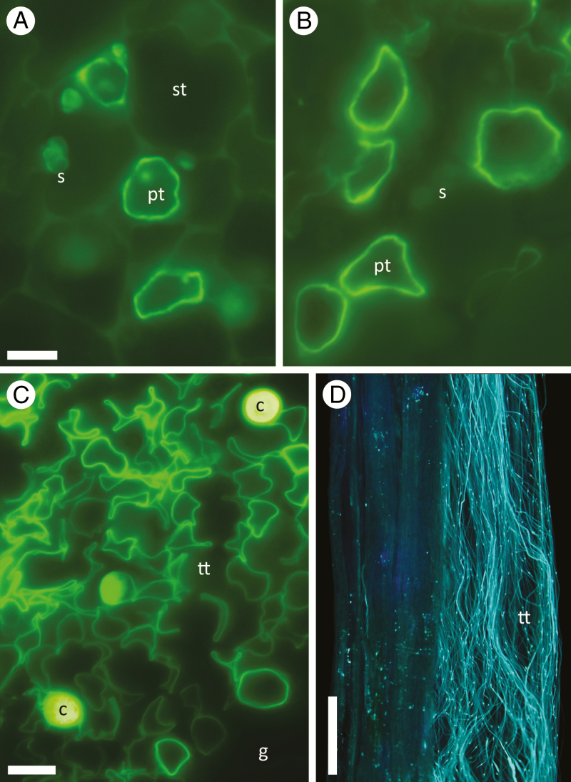
Fig. 3.
Pollen tube growth in Betula and Handroanthus. (A, B) Cross-sections of (A) haploid B. occidentalis (2x) and (B) triploid B. papyrifera (6x) pollen tubes (pt) growing between starch-bearing (s) stylar cells (st). (C) Cross-section of H. serratifolius (6x) secretory transmitting tissue (tt) showing many collapsing pollen tubes, and some callose plugs (c). Dark area is stylar ground tissue (g). (D) Longitudinal view of H. ochraceous (2x) pollen tubes with callose plugs in transmitting tissue of mid-style. All stained with aniline blue. Scale bars = A, B, 5 µm; C, 10 µm; D, 0.5 mm.

