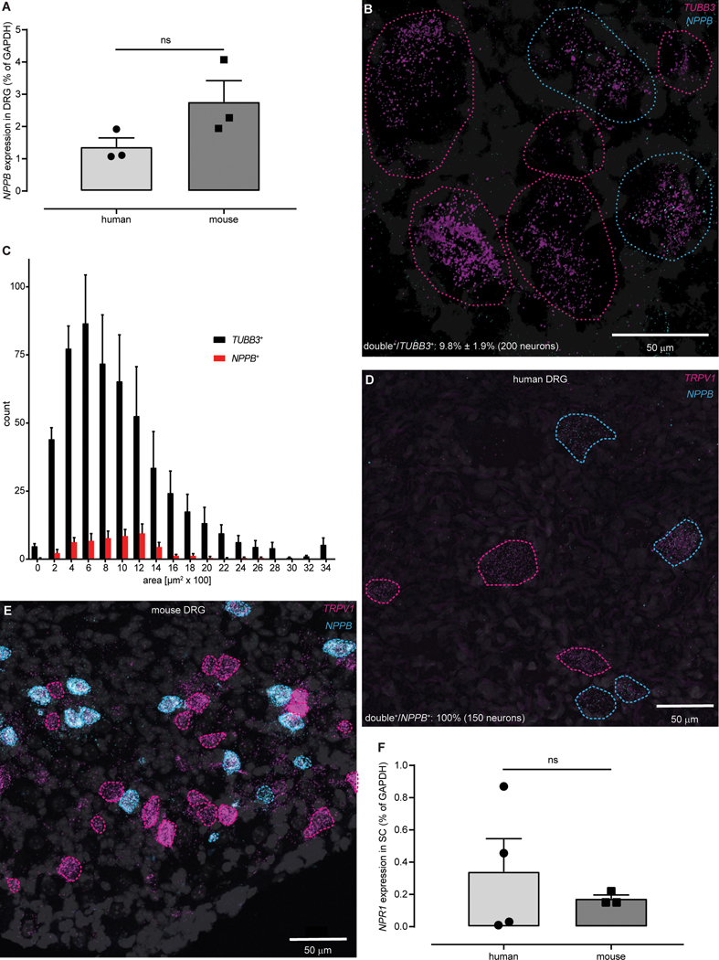
Fig. 1: The NPPB-NPR1 itch signaling pathway is conserved between mice and humans.
(A) qPCR-based quantification of expression did not show a significant difference in amounts of NPPB transcripts between human and mouse DRG (P = 0.1241, unpaired t-test, n = 3). (B) Representative double ISH images of a field of human DRG with neurons stained for NPPB (cyan) and TUBB3 (magenta). NPPB-positive and NPPB-negative neurons are outlined with cyan and magenta dots respectively, DAPI counterstain is displayed in grey. (C) Quantification of the soma-size of NPPB- (red) compared to TUBB3-stained (black) neurons (n = 4). Representative double ISH images, of fields of human (D) and mouse (E) DRG, reveal that NPPB (cyan) and TRPV1 (magenta) are co-expressed. In human and mouse DRG, NPPB is expressed in a subset of TRPV1-neurons (cyan-dotted profiles), single-labeled TRPV1-neurons are indicated with magenta-dotted profiles. (F) qPCR-based quantification of expression did not show a significant difference in amounts of NPR1 transcripts between human and mouse spinal cord (ns P > 0.9999, Mann-Whitney, n = 4 (human) 3 (mouse)).

