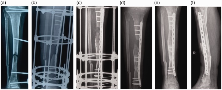
Figure 1.
Radiographs and photographs of a 47-year-old male patient (no. 8 in Table 1). (a) Before treatment, showing a 5.4-cm segmental tibial defect. (b) After placement of a laterally-based, 4.5-mm locking plate, showing a ring external fixator applied for bone transport. (c) After docking of the transport segment was achieved (after 64 days of transport). (d) After removal of the fixator on day 64, showing two percutaneous screws fixed at the transported segment. (e, f) Anteroposterior and lateral views after 9 months showing satisfactory union achieved at the distraction callus and docking site.

