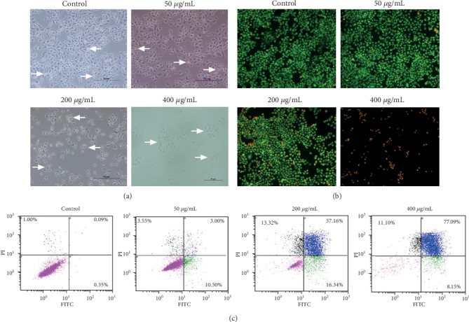
Figure 3.

Flavonoids in A. conyzoides induced significant apoptosis in HeLa cells. (a) Influences of increasing doses of flavonoids (50, 200, and 400 μg/mL) on the common morphology and density of HeLa cells were observed under the light microscope. White arrows point to the round cells (apoptosis cells). (b) Apoptosis in HeLa cells after treatments with increasing doses of flavonoids (50, 200, and 400 μg/mL) was detected by AO/EB staining. Cells with light green fluorescence in the homogeneous nuclei were considered the survival cells. Cells with bright green fluorescence and orange-red fluorescence in the pyknotic nuclei were considered the early and late apoptotic cells, respectively. (c) Apoptotic analyses in HeLa cells after treatments with increasing doses of flavonoids (50, 200, and 400 μg/mL) using a flow cytometry (black spots: necrotic cells, blue spots: late apoptotic cells, green spots: early apoptotic cells, and pink spots: survival cells).
