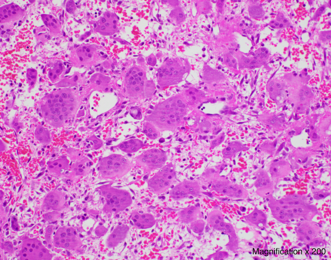
Figure 2.

Histological slice of the biopsy of the left iliac lesion showing multiple multinucleated giant cells scattered evenly in a sparse stroma, intermixed with round and spindle-shaped mononuclear cells, as well as extravasated red blood cells (Magnification× 200).

 This work is licensed under a