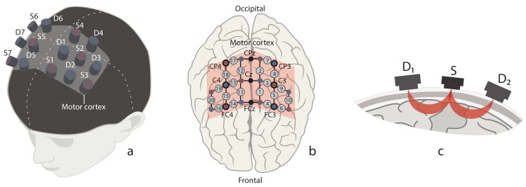
Figure 1.
(a) Location of fNIRS sources (marked with red) and detectors (marked with blue) on the subject’s head in the area of primary motor cortex (shaded area). (b) Location of 20 fNIRS channels (grey circles with channel numbers) across motor cortex (shaded area) and 9 EEG electrodes (CP4, CPz, CP3, C4, Cz, C3, FC4, FCz, FC3) according to “10–10” scheme (black circles with channel names). (c) Schematic illustration of traveling path of near-infrared light from source (S) to neighbour detectors ( and ) through brain cortex matter.

