Figure 12.
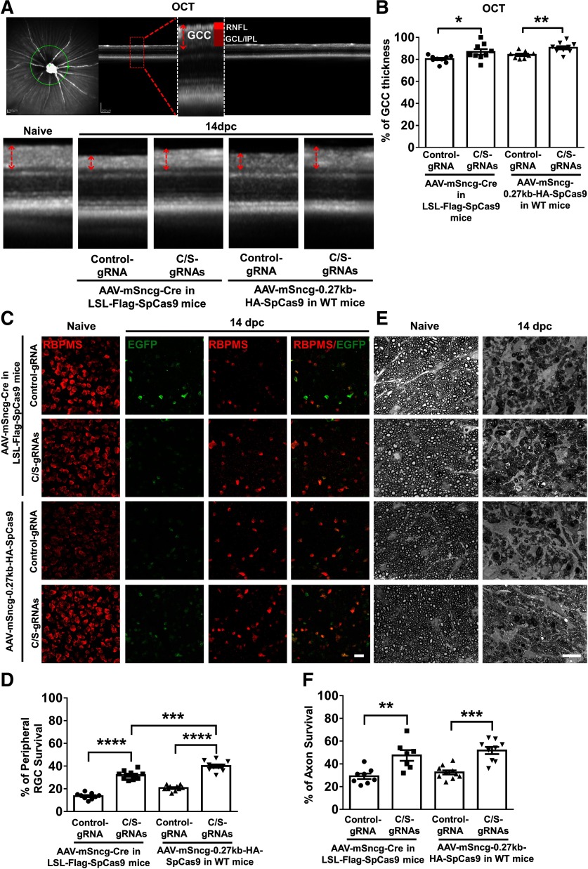
AAV-mSncg-CRISPR/Cas9 mediated Ddit3/Sarm1 inhibition promotes significant RGC soma and axon survival after ON crush injury. A, Representative OCT images of mouse retina. Green circle: indicator of the ring scanned circumpapillary region of the retina. GCC: ganglion cell complex, including RNFL, GCL, and IPL layers. B, Quantification of GCC thickness, represented as percentage of GCC thickness in the injured eyes, compared with the intact contralateral eye. Data are presented as mean ± SEM, n = 8–10; *p < 0.05, **p < 0.01, Student's t test. C, Confocal images of wholemount retinas showing surviving EGFP positive (gRNA expressed cells) and RBPMS-positive (red) RGCs, Scale bar, 20 µm. AAVs were injected intravitreally two weeks before ON crush and mice were killed 14 dpc. E, Light microscope images of semi-thin transverse sections of ON with PPD staining. Scale bar, 10 µm. D, F, Quantification of surviving RGC somata and axons, represented as percentage of crush injured eyes compared with the sham contralateral control eyes. Data are presented as mean ± SEM and n = 7–10; **p < 0.01, ***p < 0.001, ****p < 0.0001; one-way ANOVA with Tukey's post hoc test.

