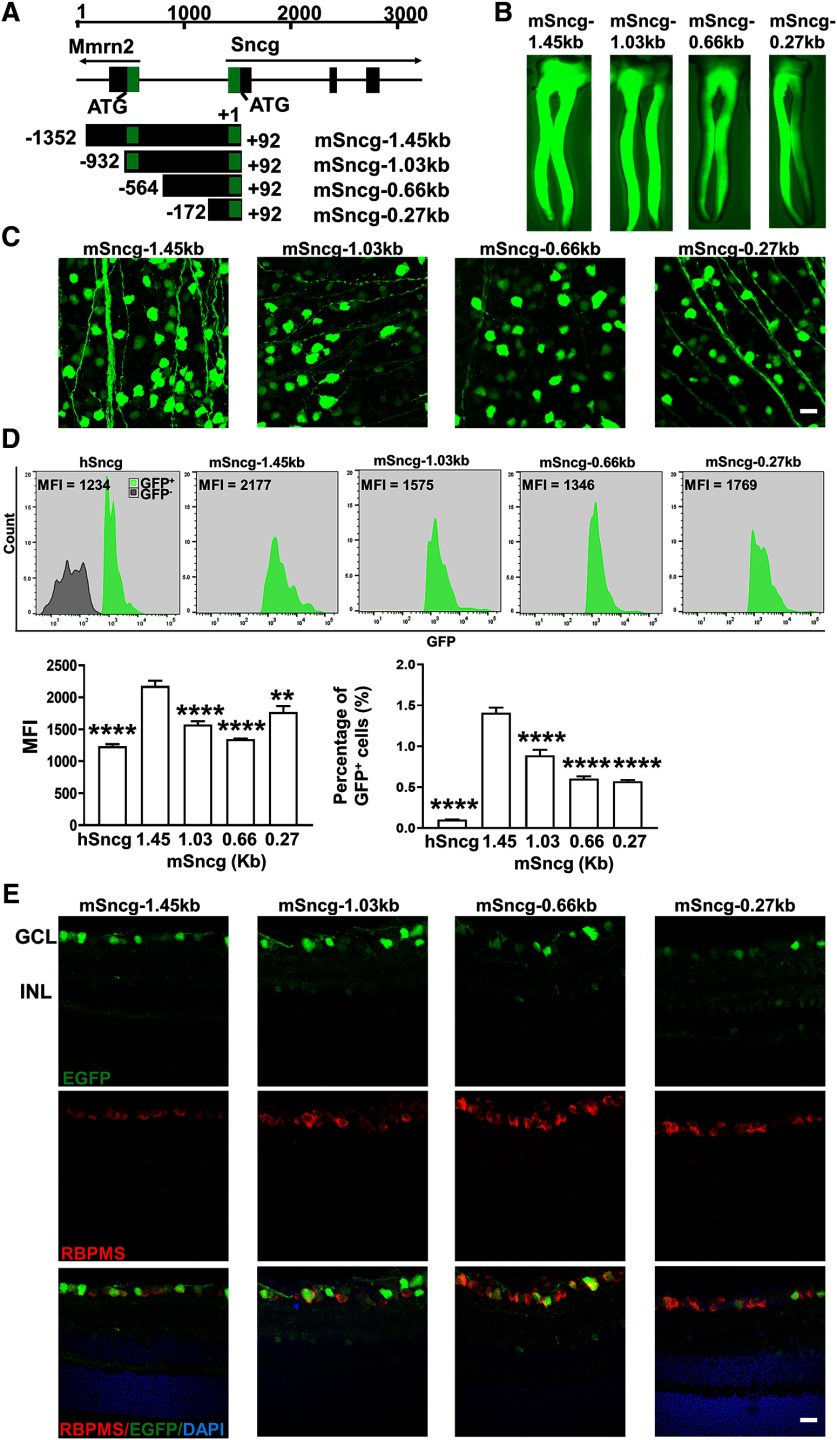
Figure 6.
The specificity and potency of different truncated forms of mSncg promoter in mouse RGCs. A, Schematic illustration of the mouse Sncg gene and its promoters. The Sncg gene is located on chromosome 14 (34370274–34374669) adjacent to multimerine 2 gene (Mmrn2). The 1.45-kb mSncg promoter starts from −1352 bp upstream of +1 Sncg transcription start site and ends at +92 bp. It includes partial sequence of the first intron and the whole first exon of Mmrn2, the non- transcription regions between Mmrn2 and mSncg genes, and the non-translated region of the first exon of mSncg. The green “boxes” represent non-translated regions. B, Epi-fluorescence low-magnification images of ON wholemounts showing EGFP expression in RGC axons. C, Confocal images of wholemount retinas showing EGFP-positive RGCs. Scale bar, 20 µm. D, The MFI and percentage of GFP+ cells of each promoter-driven GFP among total retina cells were measured by FACS. Data are presented as mean ± SEM, n = 4; **p < 0.01, ****p < 0.0001; one-way ANOVA with Tukey's post hoc test. E, Confocal images of cross sections of retinas showing EGFP signals in different retina layers. Scale bar, 20 µm.

