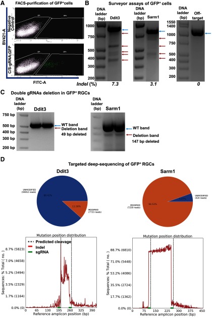
Figure 9.
The efficiencies of Ddit3 and Sarm1 genomic editing in mouse retina by AAV-mediated CRISPR/Cas9. AAV-Sncg-Cas9 and AAV-U6-C/S gRNAs-Syn1-GFP were co-injected intravitreally. A, Representative FACS plots of dissociated retinal cells gated for GFP+ cell isolation. BV421 (405 nm violet laser channel) was used to gate autofluorescence and FITC channel was used to gate GFP+ cells. A total of 10 WT mice were intravitreally injected with 6 × 10⁹ vg AAV-mSncg-Cas9 and 3 × 10⁹ vg AAV-C/S gRNAs-Syn1-GFP. B, Surveyor assay revealing indel formation at the Ddit3 and Sarm1 genomic locus, but not the off-target locus. Blue arrows are WT bands; red arrows are indel bands. C, Representative gel images showing WT and deletion bands of Ddit3 and Sarm1. Blue arrows are WT bands; red arrows are deletion bands. D, Schematic illustration of the insertion/deletion ratio and position distribution in the targeted regions of C/S gRNAs in mouse Ddit3 and Sarm1 locus, detected by deep sequencing.

