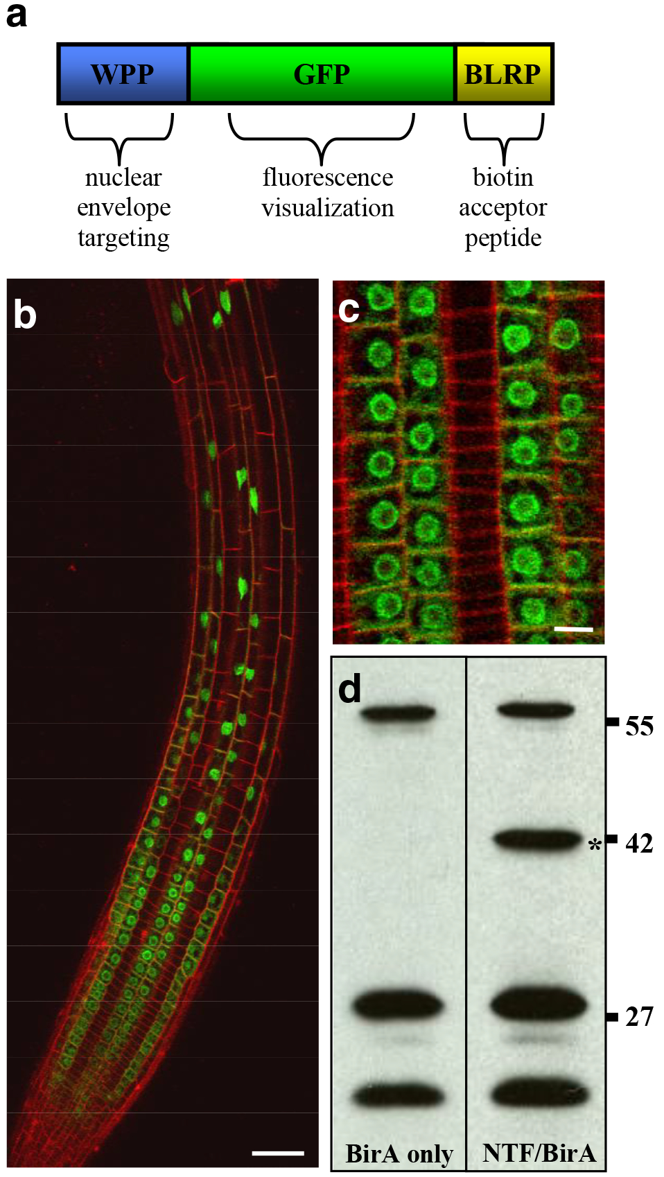
Figure 1. Nuclear targeting fusion (NTF) protein and transgenic lines for the INTACT system.

(a) The three-part structure of the NTF is shown. The chimeric protein consists of the WPP domain of RanGAP1 for nuclear envelope targeting, green fluorescent protein (GFP) to allow visualization, and the biotin ligase recognition peptide (BLRP), which is biotinylated by BirA. (b) Confocal projection image of an Arabidopsis root expressing the NTF in the epidermal non-hair cells. GFP is shown in green and cell walls are shown in red. Scale bar is approximately 20 μm. (c) Confocal section of a root expressing the NTF in non-hair cells, showing localization to the nuclear envelope. Scale bar is approximately 5 μm. (d) Streptavidin western blots of protein extracts from plants expressing BirA only or both the NTF and BirA. Asterisk indicates the position of the 42 kDa NTF. Bands found in both BirA only and NTF/BirA protein extracts are endogenous biotinylated proteins. Molecular weights (in kDa) are indicated on the right.
