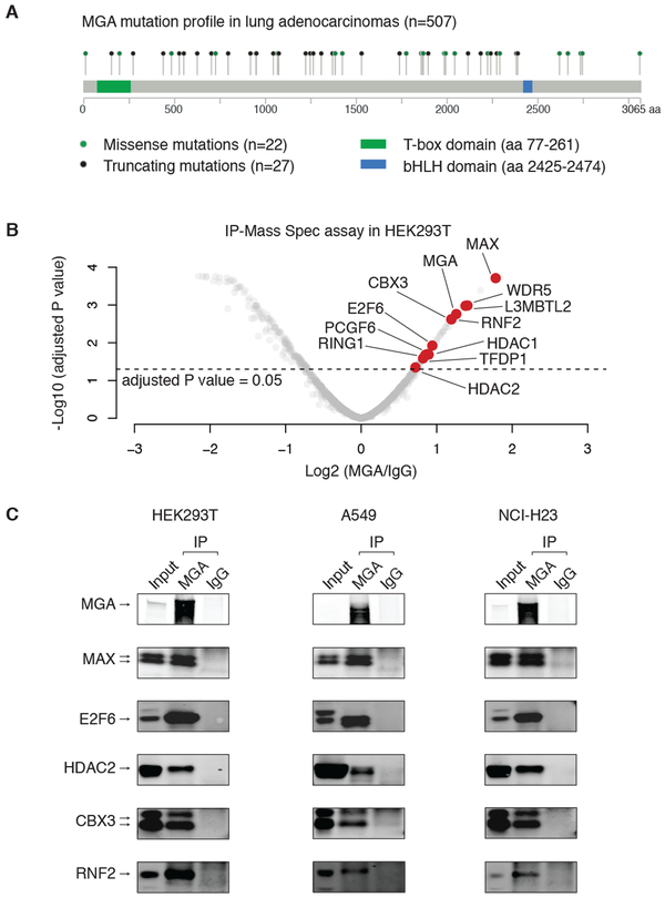
Figure 1: Genomic and proteomic analysis of MGA in lung adenocarcinomas.
A. Mutation profile of MGA in lung adenocarcinomas. Missense (green dots) and truncating mutations (black dots) are highlighted.
B. Immunoprecipitation-mass spectrometry (IP-Mass Spec) results in HEK293T cells (2 biological replicates). We identified proteins that are significantly enriched by the endogenous MGA antibody, relative to IgG control (adjusted P value < 0.05). The proteins that are part of a noncanonical PRC1 (ncPRC1) – the PCGF6-PRC1 complex – are highlighted by red dots.
C. Immunoprecipitation (IP) of MGA and IgG, followed by immunoblotting of MGA-interacting proteins in HEK293T, A549 and NCI-H23 cells.

