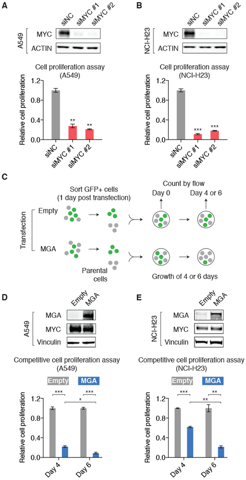
Figure 4: MGA acts as a repressor for cancer cell proliferation.
A. siRNA-mediated silencing of MYC (verified by immunoblots) decreased the proliferation of A549 cells. Cell number was counted five days post transfection and normalized to the negative control siRNA. P values are derived from t tests. Error bars: s.d., **P<0.01.
B. Same experiment as A, but in NCI-H23 cells, Error bars: s.d., ***P<0.001.
C. Schematic chart explaining the design of competitive cell proliferation assays. A549 and NCI-H23 cells were first transfected with empty-ZsGreen control or MGA-ZsGreen vector. The transfected cells were collected based on GFP signal and mixed with parental cells at 1:1 ratio. The percentage of GFP-positive cells was then counted over time.
D. MGA overexpression (verified by immunoblots) decreased the proliferation of A549 cells. The percentage of GFP-positive cells was counted four and six days post seeding the cells and normalized to the empty-ZsGreen control. P values are derived from t tests. Error bars: s.d., *P<0.05; ***P<0.001.
E. Same experiment as D, but in NCI-H23 cells, Error bars: s.d., **P<0.01; ***P<0.001.

