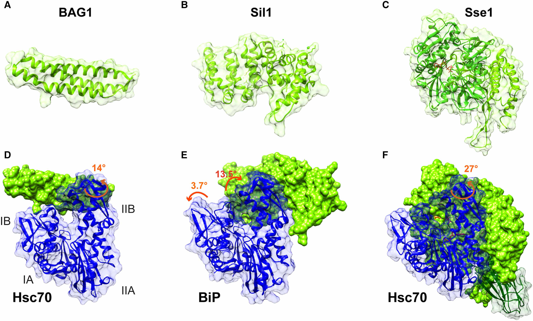
Figure 6. Structural features of eukaryotic NEFs.

Structures of human BAG1 (A), yeast Sil1 (B), and the yeast Hsp110, Sse1 (C). Crystal structures of the complexes of these NEFs with their respective Hsp70s are shown below each isolated NEF structure, with an indication of what changes in the NBD conformation are caused by NEF binding: (D) bovine Hsc70 NBD-human BAG1 (PDB ID 1hx1 [111]); (E) yeast BiP NBD-Sil1 (PDB ID 3qml [71]); (F) yeast Hsc70–Hsp110 (PDB ID 3c7n [138]). The structure shown in (F) of the complex between Hsc70 and Hsp110 includes the SBD (shown in transparent surface and green ribbon). In (F), ADP*BeF3 bound to Hsp110 and ADP bound to Hsc70 are colored in red.
