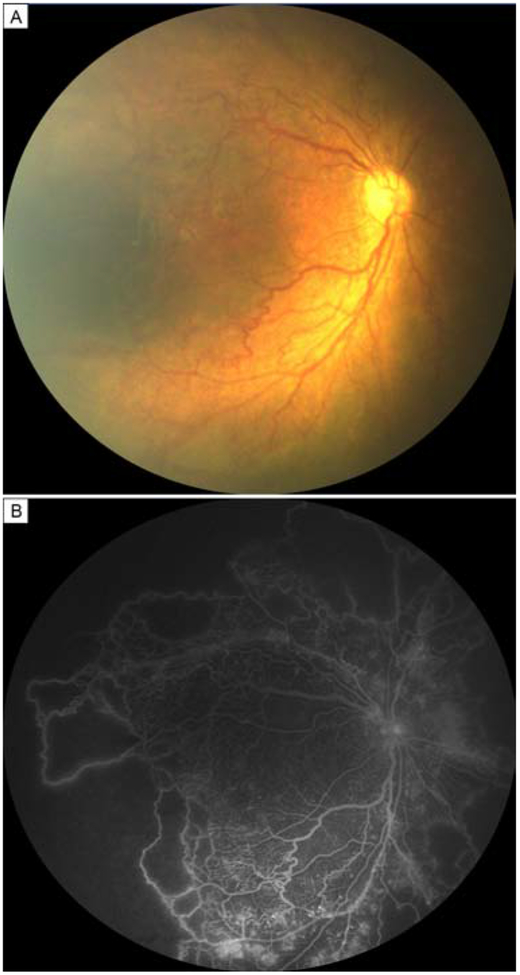
FIG 1.
Patient diagnosed with aggressive posterior retinopathy of prematurity. Fundus photograph (A) and fluorescein angiogram (B) of a 3-week-old boy demonstrating posterior disease, nonperfusion, and vascular dilation and tortuosity. The boy was born at 31 weeks’ postmenstrual age, with a birthweight of 1418 g.

