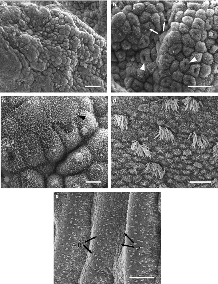
Figure 3.

Scanning electron micrographs of the uterine surface of Monodelphis domestica at Stage 2 of pregnancy (11 days 1 hr–12 days 2 hr post‐copulation). (a) Uterine region consisting of cells with domed apices. (b) Higher magnification of (a): depicts apical ‘dimples’ (arrowheads) and clumps of glycocalyx (arrows). (c) Cells with elongated microvilli (arrowhead) occur. (d) Uterine region consisting of uniform ‘honeycomb’ cells. (e) Domed uterine epithelial cells (d, arrows) are restricted to the bases of uterine folds, while flattened ‘honeycomb’ regions (h, arrows) occur on the crests. Scale bars = 25 μm (a), 20 μm (b), 5 μm (c), 10 μm (d), 100 μm (e)
