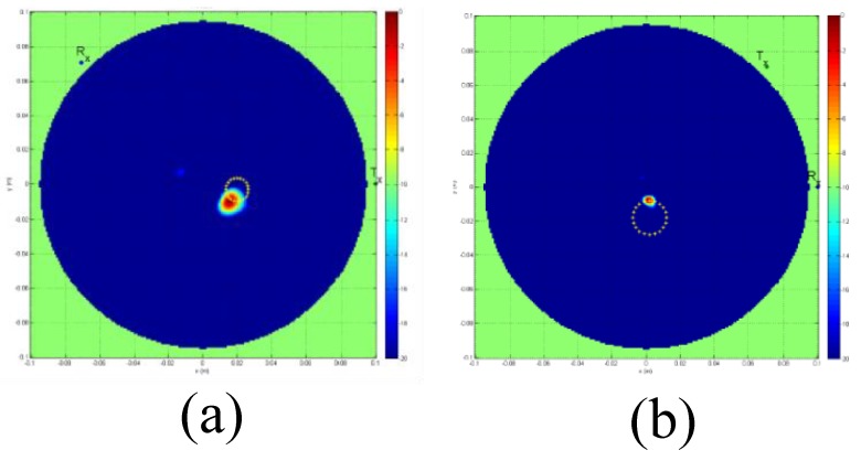
Figure 13.
Reconstructed MT images obtained using the system developed by Pagliari et al. [69]. (a) A 12 mm tumour is indicated by the red spot inside a metallic cylinder breast phantom. (b) A 12 mm tumour is indicated by the red spot inside a dielectric cylinder breast phantom. Reprinted with permission from [69].

