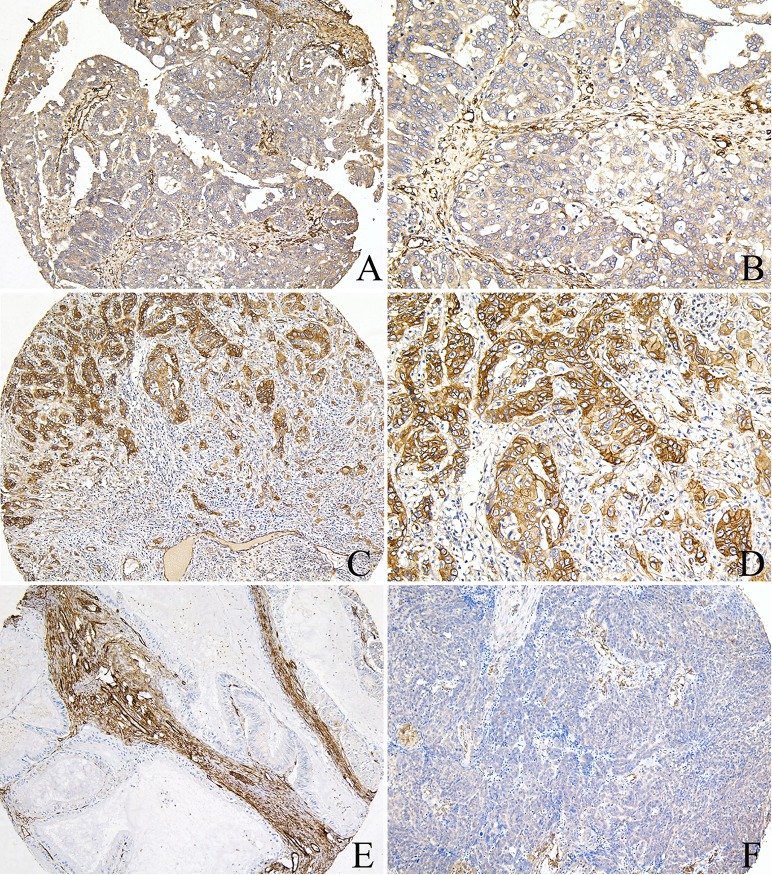
Fig 4. CAV1 protein expression in ovarian cancer tissues.
A, B) High expression of CAV1 in the tumor cells and fibroblasts, positive in the vascular endothelial cells of ovarian serous cancer tissues; C, D) High expression of CAV1 in the cancer cells, negative in the inflammatory cells of ovarian endometrioid cancer tissues; E) Low expression of CAV1 in the cancer cells, high expression in the fibroblasts of ovarian mucinous cancer tissues; F) Low expression of CAV1 in the cancer cells and fibroblasts, negative in the inflammatory cells, positive in the vascular endothelial cells of ovarian endometrioid cancer tissues (A, C, E, F: 100×IHC; B, D: 200×IHC). ATG4C protein was highly expressed in EOC cells, primarily localized in the cell membrane and cytoplasm, moreover, there was positive ATG4C expression in cancer-related fibroblasts (CAFs) and inflammatory cells (Fig 5). Among EOC tissues, high expression of ATG4C in cancer cells and stromal cells was 70.5% (67/95) and 31.6% (25/79) respectively, which was higher than that in noncancerous ovarian tissues.

