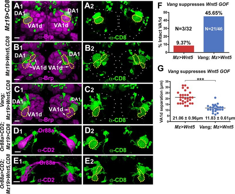
Fig 6. Vang functions downstream of Wnt5 to cell non-autonomously repel the VA1d dendrites.

(A-C) Adult ALs from animals expressing UAS-mCD8::GFP under the control of Mz19-Gal4 were stained with antibodies against Brp (magenta) to visualize the glomerular pattern and CD8 to visualize the DA1/VA1d dendrites (green). (A) In the wild-type control, the DA1 dendrites are located dorsal to the VA1d dendrites, which form a single compact arbor. (B) In animals expressing UAS-Wnt5 under the control of Mz19-Gal4, the VA1d dendrites split into two separated arbors, probably due to repulsion between the dendritic branches. (C) Removing Vang functions in animals expressing UAS-Wnt5 under the control of Mz19-Gal4 return the VA1d dendrites to its compact morphology in the right AL. In the left AL, the two separated branches are closer together. (D, E) Adult ALs from animals expressing Or88a-CD2 and UAS-mCD8::GFP under the control of Mz19-Gal4 were stained with anti-CD2 (magenta) and anti-CD8 (green) to visualize the pre- and postsynaptic processes of the VA1d glomerulus. In the wild-type control, Or88a axons are faithfully paired with the VA1d dendrites (D). In animals expressing UAS-Wnt5 under the control of Mz19-Gal4 the Or88a axons were still correctly paired with the VA1d dendrites despite their splitting into two separated arbors (E). (F) Graph summarizing the percentage of intact VA1d glomeruli in wild-type and Vang6 animals overexpressing Wnt5 in the VA1d glomeruli. (G) Graph summarizing the distance between the split VA1d arbors in wild-type and Vang6 animals overexpressing Wnt5 in the VA1d glomeruli. Student’s t test was used to compare the data of the wild-type and Vang6 animals overexpressing Wnt5. Scale bars: 10 μm.
