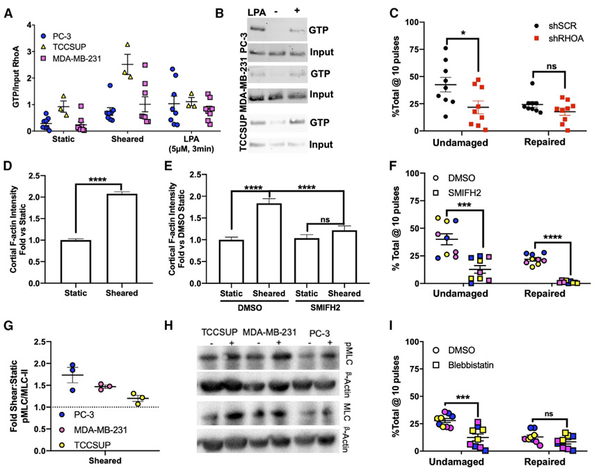
Figure 3. RhoA/Actomyosin Function Is Required for Resistance to FSS-Induced Plasma Membrane Damage.
(A) RhoA activation in PC-3 (*p < 0.05, n = 8, Welch’s t test), MDA-MB-231 (*p < 0.05, n = 8, Welch’s t test), and TCCSUP (*p < 0.05, n = 3, Welch’s t test) cells in suspension under static conditions (static), after two pulses of FSS (sheared), and in response to lysophosphatidic acid (LPA; positive control).
(B) Representative western blots of active RhoA from pull-down assay.
(C) Effects of 10 FSS pulses on the ability of cancer cells to resist plasma membrane damage (*p < 0.05, t test, n = 9) and repair damage in cells treated with control and RhoA-targeting shRNAs (p > 0.05, n = 9, t test).
(D) Effects of FSS pulses on cortical F-actin levels in PC-3 cells (****p < 0.0001, Mann-Whitney U test).
(E) Effects of SMIFH2 on F-actin levels after FSS exposure (****p < 0.0001, Kruskal-Wallis with Dunn’s correction and two-way ANOVA).
(F) Effects of SMIFH2 on the ability for PC-3, MDA-MB-231, and TCCSUP to remain undamaged and repair plasma membrane damage after 10 pulses of FSS (****p < 0.0001, t test, n = 3/cell line).
(G) Analysis of pMLC from PC-3, TCCSUP, and MDA-MB-231 after two pulses of FSS (p < 0.05 for all cell lines, t test, n = 3/cell line).
(H) Representative blot of (G).
(I) Effects of blebbistatin on membrane damage (***p < 0.01, t test, n = 3/cell line) and membrane repair (p > 0.05, t test, n = 3/cell line) in response to FSS exposure. Data in (D) and (E) are presented as median with 95% confidence interval (CI) error bars; all other data are presented as mean with SEM error bars. Original blots for data presented in (B) are found in Figure S8 with boxes to outline what is presented.
See also Figures S3-S5.

