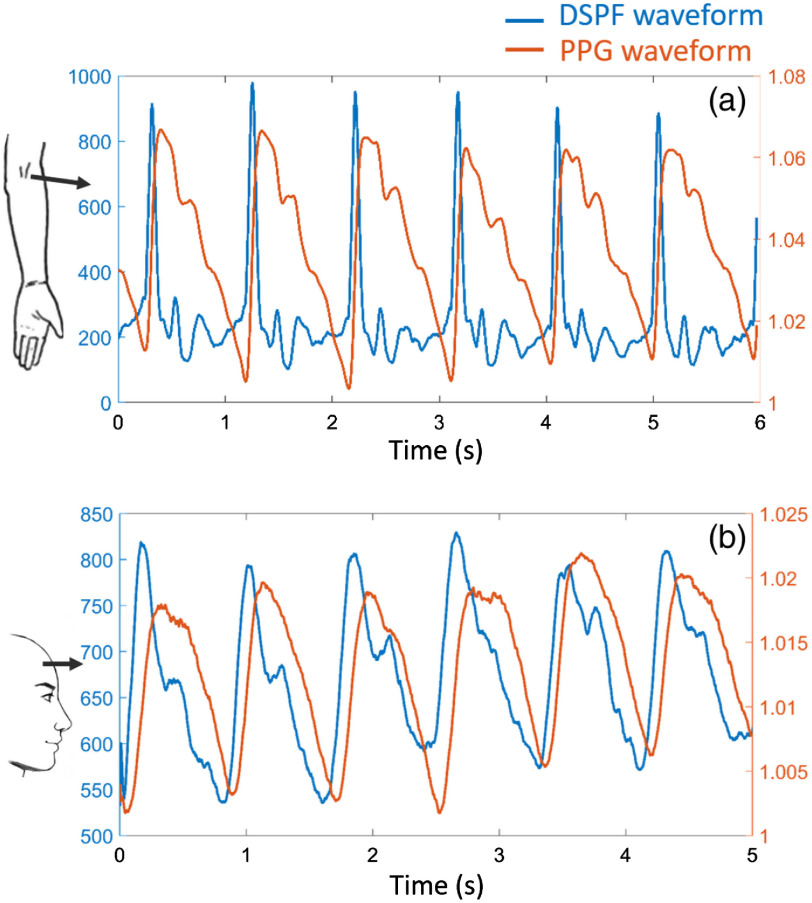
Fig. 5.
Simultaneous measurement of DSPF (blue) and PPG (orange) waveforms. Both (a) brachial artery (s–d separation: 15 mm and laser power: 4 mW) and (b) right prefrontal cortex (s–d separation: 25 mm and laser power: 20 mW) measurements are demonstrated. The exposure time of the CCD camera was 2 ms. The corresponding measurement locations are indicated on the left (Video 2, 980 KB, MP4 [URL: https://doi.org/10.1117/1.JBO.25.5.055003.2]).

