Figure 1.

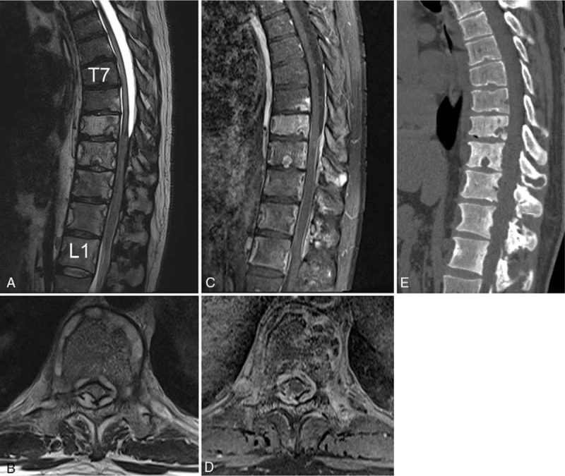
Preoperative T2-weighted (A and B), T1 gadolinium-enhanced (C and D) sagittal MR images, and axial images at the level of T10, (E) a sagittal CT reconstructive image showing a homogeneous continuous enhancing epidural lesion and osteolytic bone lesion at the T7–L1 level.
