Figure 3.

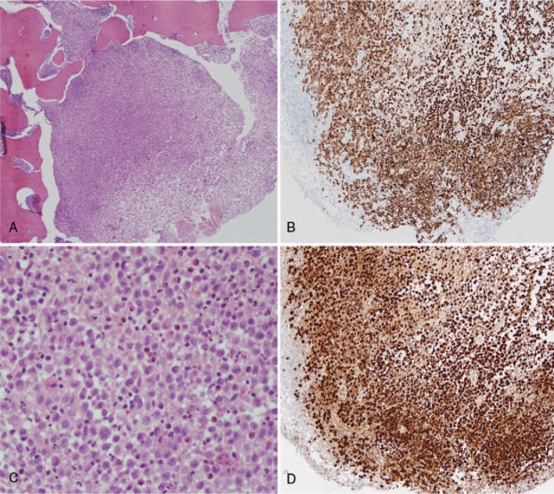
Photomicrographs of the epidural lesion. The tumor is composed of a large number of B cells and plasma cells. (A) H&E staining, X40, (B) H&E staining, X400, (C) CD1a staining, and (D) S-100 immunohistochemical staining.

Photomicrographs of the epidural lesion. The tumor is composed of a large number of B cells and plasma cells. (A) H&E staining, X40, (B) H&E staining, X400, (C) CD1a staining, and (D) S-100 immunohistochemical staining.