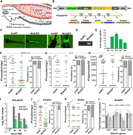
Fig. 1. Generation of transgenic mosquitoes (MultiEff) using an AgCp-driven transgene array with five anti-Plasmodium effectors targeting the malaria parasite in the midgut, and P. falciparum infection phenotypes at both oocyst and sporozoite stages.

(A) Schematic illustration of transgenic targeting of parasite midgut infection stages and the design of the transgenes to target the parasites at this stage. (B) Five known anti-Plasmodium effectors (Melittin, TP10, EPIP, Shiva1, and Scorpine) were selected. The gene array with these five anti-Plasmodium effector genes was synthesized through GenScript Inc. and cloned under the AgCp promoter, followed by the trypsin terminator (TrypT) in the pBac[3xP3-EGFPafm] vector. These five antiparasitic effector genes were transcribed on one cassette separated by viral P2A sequences, translated into one polypeptide, and self-spliced into five individual peptides with an extra viral P2A amino acid tag on the first four peptides. (C) Fluorescent images of a positive larva and an adult transgenic mosquito. (D) Polymerase chain reaction (PCR) validation of the partial [500 base pairs (bp)] transgene cassette of MultiEff in the transgenic mosquitoes. (E) Transcript abundance of the transgene in the gut of MultiEff transgenic mosquitoes at various time points post-blood meal (PBM). Each bar represents the relative fold change in the transgene as compared to the control at time 0 hour. The S7 ribosomal gene was used to normalize the complementary DNA (cDNA) templates. Error bars indicate SEM. (F to H) P. falciparum (NF54) oocyst and sporozoite infection intensities and prevalence at 8 days post-infection (dpi) in the gut or 14 dpi in the salivary glands (SG) when fed on blood with a medium (0.05%) (F) or low (0.01%) (G and H) gametocytemia. At least three biological replicates were pooled for the dot plots. Each dot represents the number of parasites in an individual gut or a pair of salivary glands, with the median values indicated by red bars. P values were calculated by a Mann-Whitney test. Detailed statistical analysis is presented in table S2. (I) Midgut microbial flora of female transgenic MultiEff and WT control (AsWT) mosquitoes at 0-, 24-, 48-, and 72-hour PBM (mean ±SEM). (J and K) P. falciparum oocyst infection intensities and prevalence in the aseptic (antibiotic-treated) and septic (non–antibiotic-treated) transgenic and AsWT mosquitoes at 8 dpi. (L) Expression of AMP and anti-Plasmodium effector as fold change in expression through quantitative reverse transcription PCR (qRT-PCR). Error bars indicate SEM. CEC1, Cecropin 1; DEF1, Defensin 1; GAM1, Gambicin 1; FBN9, Fibrinogen-related protein 9.
