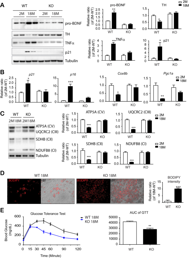
Figure 6.
PDGFRα+ cell-specific KO reduced inflammatory and senescence marker expression in eWAT and insulin resistance of mice with advanced age. (A) Immunoblot analysis of BDNF and TH expression in epididymal white adipose tissue (eWAT) of BDNFpdgfra KO and WT mice at the indicated ages. (n = 5 per condition, mean ± S.E.M, *p<0.05, **p<0.01, *** p < 0.001). (B) Quantitative PCR analysis of eWAT of BDNFpdgfra KO and WT mice at the indicated ages. (n = 5, mean ± S.E.M, *p<0.05, **p<0.01, ***p<0.001). (C) Immunoblot analysis of mitochondrial makers involved in mitochondrial oxidative phosphorylation. (D) BODIPY staining of adipocytes differentiated from PDGFRα+ cells that were isolated from eWAT of BDNFpdgfra KO and WT mice (n = 4, mean ± S.E.M, **p<0.01). (E) Measurement of glucose tolerance test (GTT) in WT and BDNFpdgfraKO mice and the area under the curve of GTT plots. N = 5, mean ± S.E.M, **p<0.01. Full images of Western blots are shown in supplementary Fig. 6.

