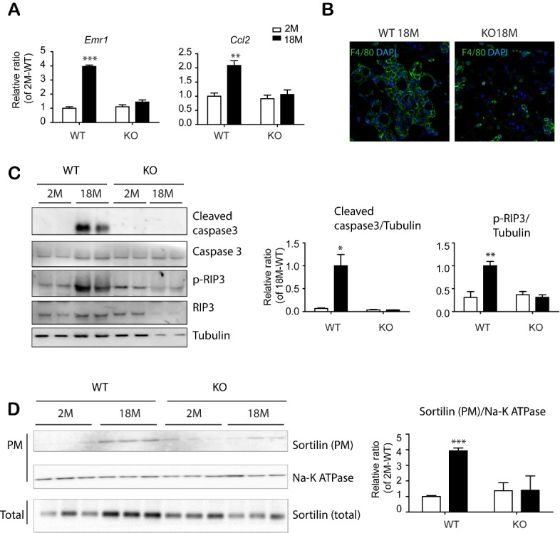
Figure 7.

PDGFRα+ cell-specific KO reduced apoptosis and necroptosis in eWAT of mice with advanced age. (A) Quantitative PCR analysis of eWAT of BDNFpdgfra KO and WT mice at the indicated ages. (n = 5, mean ± S.E.M, *p<0.05, **p<0.01, ***p<0.001). (B) Immunostaining of F4/80 in paraffin sections of eWAT of BDNFpdgfra KO and WT mice. DAPI was used as a nuclear counterstain. (C) Immunoblot analysis of apoptosis/necroptosis makers in eWAT of BDNFpdgfra KO and WT mice. (D) Immunoblot analysis of sortilin expression in plasma membrane fractions of eWAT of WT and BDNFpdgfra KO mice (n = 5, means ± SEM). Full images of Western blots are shown in supplementary Fig. 7.
