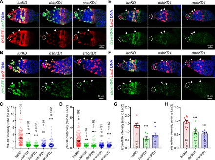
Fig. 1. Hh and Wnt signaling are interdependent in the differentiation niche.

The germaria are labeled for PZ1444-LacZ expression to visualize IGS cells (two indicated by arrowheads) and cap cells (broken ovals), while DAPI staining identifies all nuclei. (A to D) Merged confocal images of germaria showing that the expression of both fz3-RFP (A) and ptc-GFP (B) is significantly decreased in smoKD and dshKD IGS cells 2 days after knocking down compared with the control (lucKD) (C and D) quantification results on fz3-RFP or ptc-GFP intensities normalized to LacZ in IGS cells, respectively; n = IGS cells number. (E to H) Merged FISH (green) and immunostaining (LacZ, red) confocal images showing that fz3 (E) or ptc (F) mRNA expression levels are significantly reduced in dshKD and smoKD IGS cells (G and H: quantification results on fz3 and ptc mRNA levels based on the fluorescence intensities normalized to LacZ, respectively; n = germarial number). Scale bars, 10 μm (all images at the same scale). In this study, all the quantitative data are shown as means ± SEM, whereas P values are determined by the two-sided Student’s t test (***P ≤ 0.001; **P ≤ 0.01).
