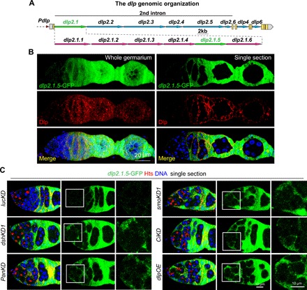
Fig. 4. Canonical Hh and Wnt signaling repress dlp transcription via the dlp2.1.5 genomic region in the second intron.

(A) Diagram of the dlp genomic regions showing dlp2.1 and dlp2.1.5 regions driving GFP expression, which recapitulates dlp mRNA and protein expression in the germarium (please see fig. S5 for details). (B) Immunostaining with anti-GFP (green) and anti-Dlp (red) showing dlp2.1.5-GFP has similar expression pattern with endogenous Dlp, which has very low level at IGS cells but high level at late-stage somatic cells. (C) Single confocal cross-sectional images of germaria showing that dlp2.1.5-GFP expression is up-regulated in dshKD1, smoKD1, panKD, ciKD, and dlpOE IGS cells compared with the control (lucKD) (anterior germarial regions highlighted by squares are shown at a high magnification). Scale bars, 20 μm in (B), 10 μm in (C).
