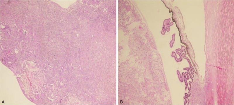
Figure 4.

Histopathology in advanced Coats’ disease. (A) Photomicrograph showing typical proteinaceous SRF with cholesterol clefts in an eye enucleated for advanced Coats’ disease (Original magnification ·100, hematoxylin and eosin). (B) Photomicrograph of anterior chamber and the ciliary body showing cholesterol clefts (Original magnification ·100, hematoxylin and eosin), and there were no tumor cells suggestive of Retinoblastoma. SRF = subretinal fluid.
