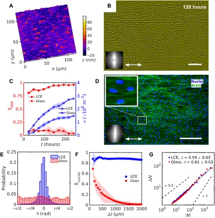
Fig. 1. Uniform alignment of HDF cells on LCE with a uniform .

(A) Digital holographic microscopy (DHM) texture of the LCE surface after contact with the aqueous growth medium. (B) Phase contrast-microscopy (PCM) texture of HDF cells growing on LCE substrates at 120 hours after seeding. Double-headed arrow represents . (C) Evolution of the order parameter SHDF of cells bodies (filled red symbols) and cell density σ (empty blue symbols). (D) Fluorescent microscopic textures of HDF cells on LCE; fluorescently labeled nuclei (blue) and cytoskeleton F-actin proteins (green). Magnified texture shows elongated nuclei oriented in the same direction as the cells’ bodies. Insets in (B) and (D) show fast Fourier transformation of (B) PCM and (D) fluorescent F-actin textures indicating orientational order along the uniform . (E) Distribution of nuclei orientation. (F) Dependence of the order parameter Snuclei of nuclei on the size of a square subwindow. (G) Number density fluctuations ΔN calculated for the mean number of cell nuclei 〈N〉. Scale bars, 300 μm.
