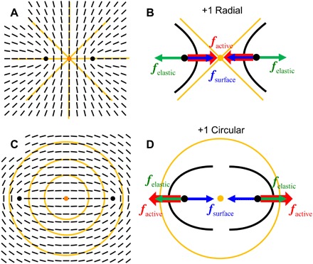
Fig. 5. Schemes of defect splitting and dynamics in patterned tissue of HDF cells.

(A) A radial +1 defect in the underlying (orange lines) and a corresponding split pair of +1/2 defects in the HDF tissue, the cells in which follow shown as black bars. (B) Splitting of the two +1/2 defects in the radial configuration is favored by elasticity (green arrow forces) of the tissue but opposed by its surface anchoring (blue arrows) and by activity (red arrows). (C and D) Similar diagrams for a circular +1 defect; in this case, the active force assists elastic repulsion in splitting, thus producing a larger separation of the +1/2 defects.
