Figure 2.

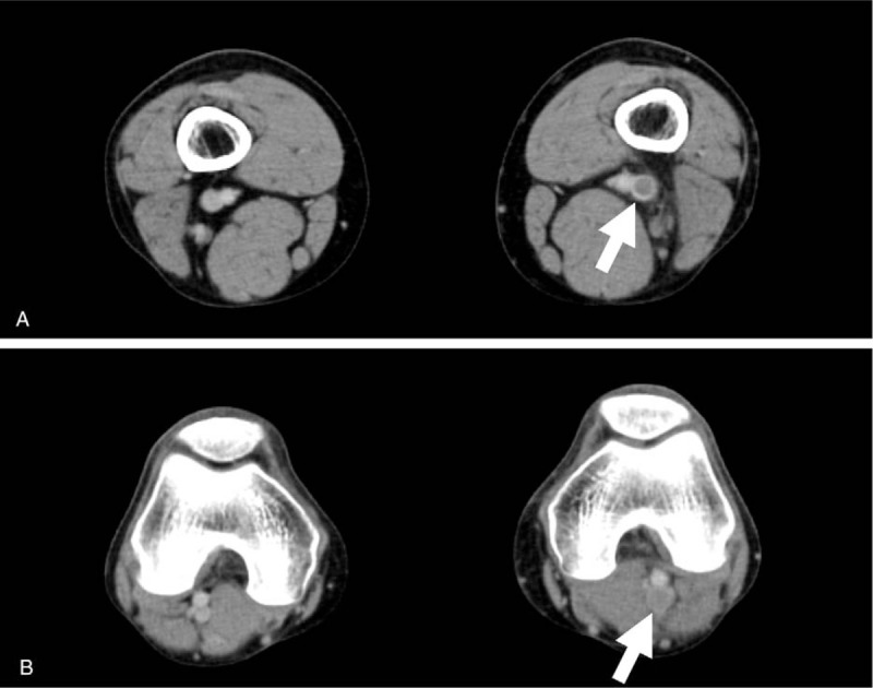
Contrast enhanced computed tomography on admission showing a thrombus from left femoral vein (A) to the left popliteal vein (B) (arrows).

Contrast enhanced computed tomography on admission showing a thrombus from left femoral vein (A) to the left popliteal vein (B) (arrows).