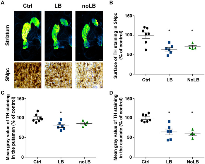
Fig. 2. Intrastriatal injection of LB and noLB fractions from patients with PD induces nigrostriatal neurodegeneration in baboon monkeys.

(A) TH staining at striatum and SNpc levels. A green fire blue LUT (lookup table) was used to enhance contrast and highlight the difference between noninjected and LB- and noLB-injected baboon monkeys at the striatum level. Scale bars: 5 mm (striatum) and 10 μm (SNpc). (B) Scatter plot of TH immunostaining in SNpc (F2,14 = 9.439, P = 0.0025; control versus LB-injected, P = 0.0029; control versus noLB-injected, P = 0.0248). (C and D) Scatter plots of mean gray values of striatal TH immunoreactivity in the putamen (F2,14 = 7.313, P = 0.0067; control versus LB-injected, P = 0.0059) (C) and in the caudate (F2,14 = 16.25, P = 0.0002; control versus LB-injected, P = 0.0008; control versus noLB-injected, P = 0.0008) (D) in noninjected and LB- and noLB-injected baboon monkeys. The horizontal line indicates the average value per group ± SEM (n = 7 from control animals; n = 6 for LB-injected animals; n = 4 for noLB-injected animals). Comparison was made using one-way ANOVA and Tukey’s correction for multiple comparisons. *P < 0.05 compared with control animals.
