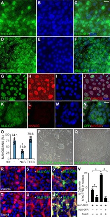
Fig. 7. Nuclear localization of TFE3 is critical for primed to naïve conversion.

(A to F) Primed H9 hESCs stably expressing TFE3-GFP fusion proteins (A to C) or NLS-mutated TFE3-GFP (NLS-GFP) (D to F) were costained for GFP (A and D) and DAPI (B and E). (G to Q) Primed H9 hESCs stably expressing TFE3-GFP (G to J) or NLS-GFP (K to N) went through the conversion protocol in Fig. 1J and were stained at day 5 for GFP (G and K), NANOG (H and L), and hNA (I and M). Merged images (J and N) were quantified for the percentage of NANOG+ cells in human nuclear antigen (hNA+) cells (O). *P < 0.05, n = 4, one-way ANOVA versus control H9. Phase-contrast images of H9 expressing TFE3-GFP (P) or NLS-GFP (Q) were acquired at day 5 of conversion. Scale bars, 10 μm. (R to V) HEK293 cells transfected with MYC-TFE3 alone (R and T) or together with NLS-GFP (S and U′) were treated with vehicle (R to S′) or Torin1 (10 μM for 3 hours) (T to U′) and stained as indicated. Merged images (S′ and U′) highlighted cells transfected with NLS-GFP. Percentage of cells with MYC-TFE3 in nucleus was quantified for each condition. *P < 0.05, Student’s t test, n = 250 cells per condition. Scale bars, 10 μm.
