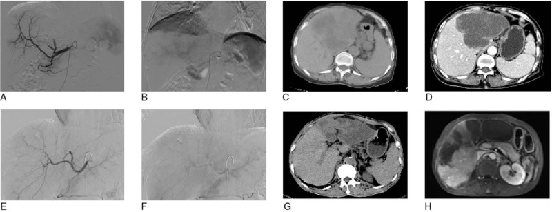
Figure 3.

CT and DSA images of 2 patients at pre-operation and post-operation. (A–D) CT and DSA images of 1 patient at preoperation and postoperation. (E–H) CT and DSA images of another patient at preoperation and postoperation. Scan parameters were that: tube voltage was 120 kv; automatic pipe technology to adjust current; collimator width was 128 × 0. 625 mm; the reconstruction was 5 mm; a thick layer of reconstruction interval was 5 mm; screw pitch was 0.914. Enhanced scan USED MEDRAD double cylinder of high-pressure syringe injected iodine alcohol (containing iodine 300 mg/ml) from peripheral vein intravenous (dose of 1.5 ml/kg). When abdominal aorta CT value arrive to 100 HU, it delays after 5 seconds and begins to scan, and after arterial phase late 30 seconds and 150 seconds, respectively, it began to scan portal venous phase and delayed phase.
