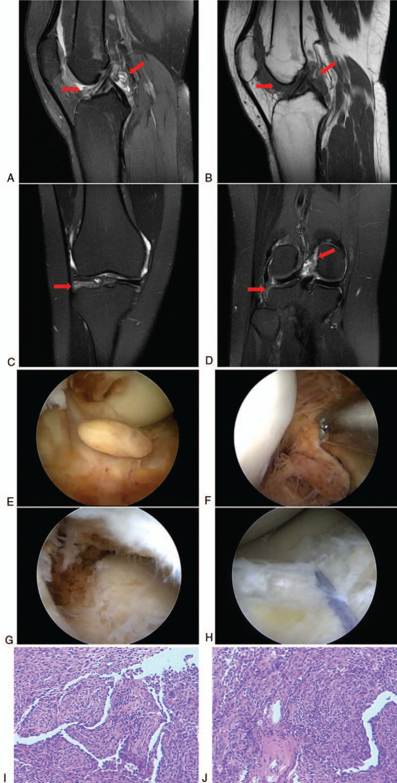
Figure 3.

Follow-up MRI performed 18 mo after the initial surgery. (A) Sagittal MRI T2WI sequence and (B) sagittal MRI T1WI sequence showed the intra-articular recurrent lesions (arrows). (C, D) Coronal MRI T2WI sequence showed the intra-articular recurrent lesions (arrows). Arthroscopic synovectomy was performed in the second surgery. (E) Intraoperative arthroscopic picture demonstrating synovial proliferation suggestive of recurrent pigmented villonodular synovitis. (F) Intraoperative arthroscopic picture demonstrating the recurrent lesion in posterior compartment of the knee. (G) Intraoperative arthroscopic picture demonstrating the recurrent lesion under the meniscus. (H) Intraoperative arthroscopic picture demonstrating that the meniscus was repaired after resection of the lesion. Pathological examination of the tissues excised in the second surgery. (I, J) Hematoxylin and eosin staining revealed hypertrophic synovium typical of pigmented villonodular synovitis. MRI = magnetic resonance imaging.
