
Figure 1.
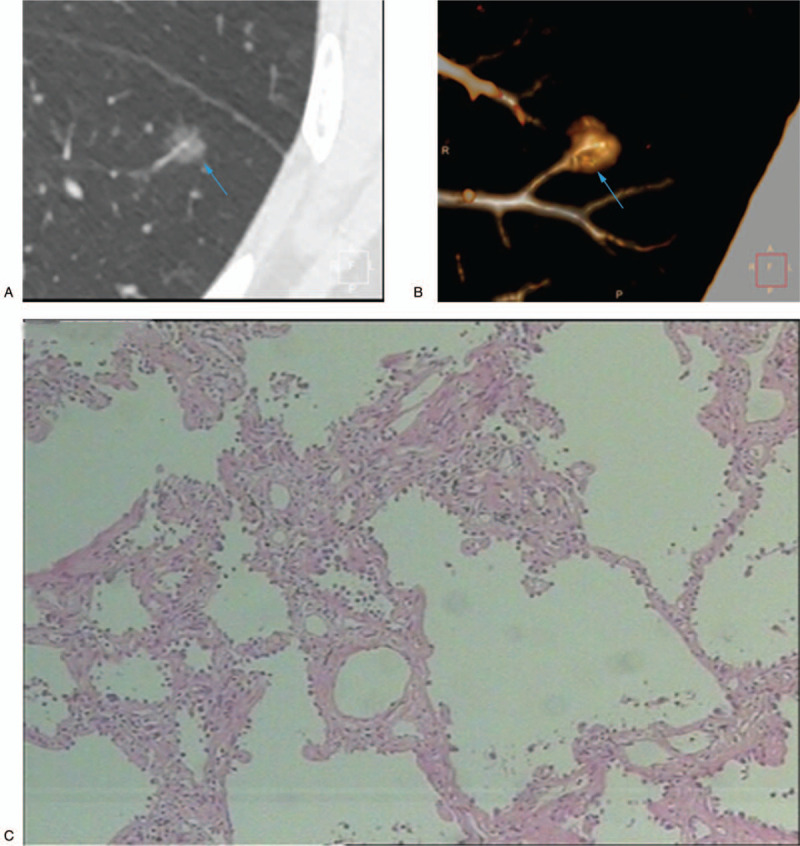
A 42-year-old nonsmoking woman with a peripheral pure ground-glass nodule in the left lower lobe. (A) High-resolution computed tomography showed a 7-mm nodule with the vascular penetration sign (blue arrow). (B) Three-dimensional reconstruction image of the same nodule (blue arrow). (C) The pathologic diagnosis of the resected specimen showed adenocarcinoma in situ.