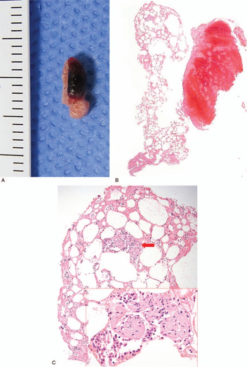
Figure 4.

Gross and microscopic findings in an old man (case 2). (A) The aspirated material from the distal basilar artery shows an 8-mm-sized lump of yellowish adipose tissue with an attached blood clot. (B) The specimen shows morphologic correlation on a low magnification image. (C) Variably sized mature adipocytes are intermixed with fibrin and blood cells and surrounding a small fragment of entrapped soft tissue (red arrow). An enlarged image of the fragment reveals a histology resembling vessel wall.
