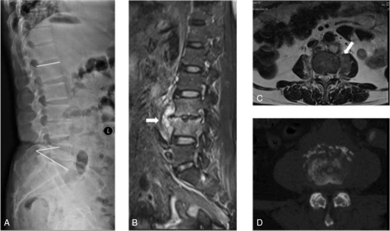
Figure 1.

. Preoperative imaging of a 62-year-old woman with vertebral osteomyelitis caused by Escherichia coli and Mycobacterium tuberculosis (case 1 in Table 1). A 62-year-old female patient with L3/4 spondylodiscitis and osteitis. Preoperative neurologic impairment was Frankel grade D. Preoperative X-ray (A) and computed tomography (D) scan show L3/4 bone destruction, and the Kyphosis angle is 35°. MRI (fat-suppressed, gadolinium-enhanced, T2-weighted MRI scan, panels B and C) shows lesions with paravertebral abscess. MRI scan shows bone marrow and disk edema, spinal epidural abscess (the white arrow in panel B), disk enhancement, and a small prevertebral abscess (the white arrow in panels B and C). MRI = magnetic resonance imaging.
