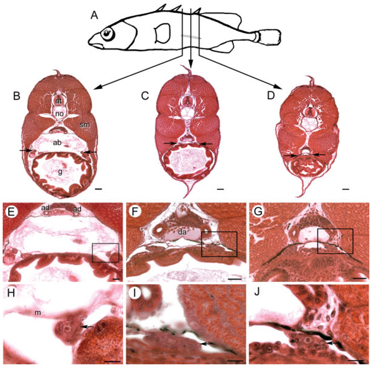
Fig. 3.
Anatomical orientation of developing threespine stickleback (Gasterosteus aculeatus) gonads at 15 dpf. A: Vertical lines, about 540 μm apart depict locations of transverse sections through the outlined left gonad of a female fry sampled from the Bear Paw population. B–D: Transverse sections with arrows showing location of the gonads. E–G: Higher magnifications show the relationship of the gonad to the mesentery (m) and archinephric duct/mesonephros. H–I: Boxed regions of above sections emphasize that gonads (arrows) flatten and approach the midline toward the posterior aspect. ab, air bladder; ad, archinephric duct; da, dorsal aorta; g, gut; m, mesentery; no, notochord; nt, neural tube; sm, skeletal muscle. Scale bars (B–D): 50 μm; (E–G): 25 μm; (I–K): 10 μm.

