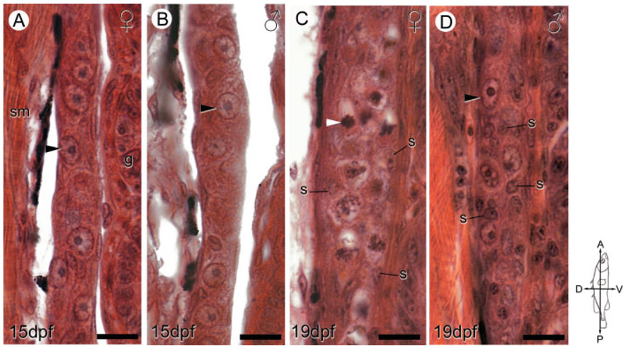
Fig. 4.
Sex-specific ovarian and testicular differentiation between 15 and 19 dpf. Representative sagittal sections of genetically female (A, C) and male (B, D) anadromous threespine stickleback (Gasterosteus aculeatus) fry at 15 dpf (A, B) and 19 dpf (C, D) highlight the dimorphic distribution of gonadal somatic (s) cells and onset of meiosis in female germ cells (C, white arrowheads). Female fry display peripheral distribution of somatic cells (C), while male fry possess many somatic cells in the gonad interior (D). The compass denotes the orientation of the fry. Black arrowheads show primordial (premeiotic) germ cells. g, gut; s, somatic cell nuclei; sm, skeletal muscle. Scale bars: 10 μm.

