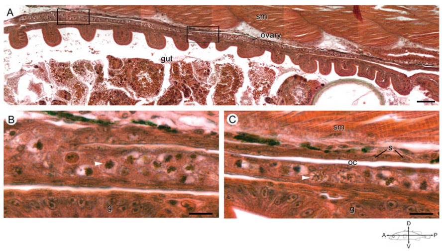
Fig. 6.
The ovarian cavity in an anadromous female threespine stickleback (Gasterosteus aculeatus) at 19 dpf. A: Photograph is a low magnification of the longitudinal axis of an ovary in a female fish sampled from the Rabbit Slough population at 19 dpf. Scale bar indicates 25 μm. Compass indicates the orientation of the fish. B, C: Magnification of the anterior (B) and posterior (C) boxed areas from (A) highlight an apparent fold in the anterior portion of the ovary forming a thin slit, ovarian cavity (oc), lined with somatic cells. White arrowheads indicate representative meiotic germ cells. Note that germ cells occur predominately on the ventral aspect of the slit. g, gut; oc, ovarian cavity; s, somatic cell nuclei; sm, skeletal muscle. Scale bar indicates 10 μm.

