Fig. 1.
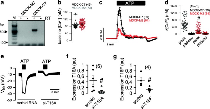
TMEM16A augments Ca2+ signaling and ion transport in MDCK cells. a RT-PCR indicating expression of TMEM16A in MDCK-C7 cells but not in MDCK-M2 cells. b Summary of basal Ca2+ levels in MDCK-C7 and MDCK-M2 cells. c, d Assessment of intracellular Ca2+ using the Ca2+ sensor Fura2. ATP or UTP (both 100 μM) increased intracellular peak and plateau Ca2+ in MDCK-C7 and MDCK-M2 cells. e Original recordings and summary of ATP or UTP induced transepithelial voltages in MDCK-C7 cells and effect of TMEM16A-knockout. f, g Effect of siRNA on expression of TMEM16A and TMEM16F, respectively, as assessed by quantitative RT-PCR. Mean ± SEM (number of cells measured). #Significant difference when compared to MDCK-C7 and scrambled, respectively

