Figure 1.

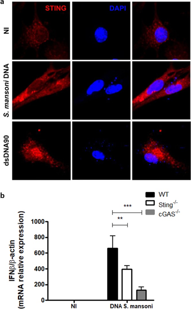
Schistosoma mansoni DNA recognition and STING activation in murine embryonic fibroblasts (MEFs). (a) Confocal microscopy of C57BL/6 (WT) MEFs stained with anti-STING (red) and DAPI (4,6-diamidino-2-phenylindole) (blue), transfected with Fugene only (NI) or transfected with 3 μg/mL of S. mansoni DNA or 3 μg/mL of STING-activating dsDNA (dsDNA90 base pairs) for 6 hours. (b) Quantitative reverse transcriptase–PCR (qRT–PCR) analysis of interferon-β (IFN-β) mRNA in WT and cGAS-/- and Sting−/− MEFs transfected with Fugene only (NI, n = 3) or transfected with 3 μg/mL of S. mansoni DNA (n = 3) for 6 hours. NI represents transfected MEFs with Fugene only. (***) and (**) are used to demonstrate statistical differences with p < 0.001 and p < 0.01 compared to the WT MEFs, respectively. Two-Way ANOVA with Bonferroni adjustments were included for multiple comparisons.
