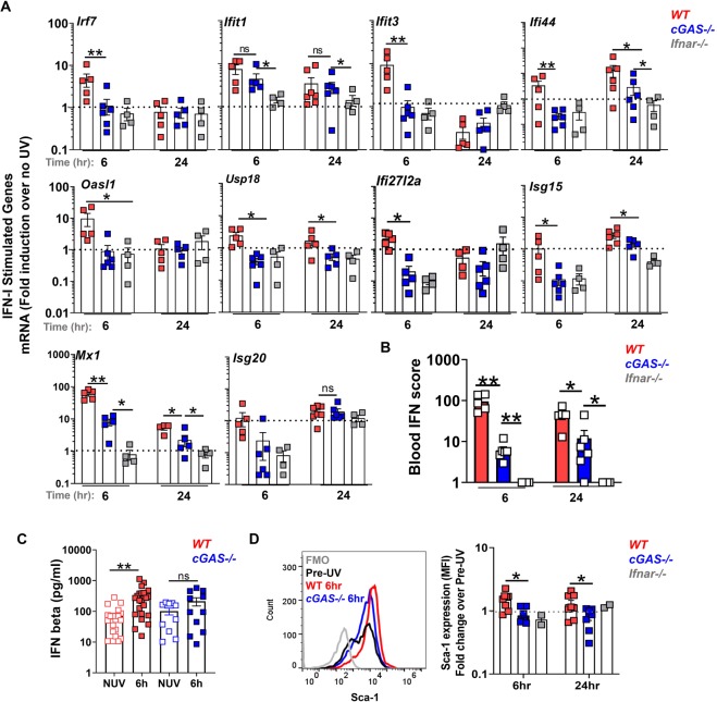
Figure 5.
cGAS contributes to the systemic IFN-I response to skin UV light exposure. Age-matched female B6 (wild type, WT), cGAS−/−, and Ifnar1−/− mice were exposed to a single dose of UVB light as in Fig. 2. (A) Fold induction in ISG mRNA levels in the peripheral blood cells 6 and 24 hr after skin exposure to UVB light was determined relative to mRNA levels in the blood prior to UV. (B) Blood IFN scores for each genotype were calculated as the sum of normalized expression levels of the 7 most highly expressed ISGs after UV exposure (Mx1, Ifit1, Ifit3, Ifi44, Usp18, Oasl1, and Ifi27l2a). (C) IFNβ concentration in plasma prior to UV (No UV, NUV) and 6 h after UV light exposure in wild-type (WT) and cGAS−/− mice. (D) Flow cytometry analysis of Sca-1 expression on B cells in the blood of wild type (WT), cGAS−/−, and Ifnar−/− mice 6 and 24 hr after UVB exposure, presented as fold change relative to non-irradiated skin cells. Representative histograms are shown for B cell population. Statistical significance was determined by Student’s t-test (n = 4–6, A,B; n = 11–23, C; n = 3–8, D; *p < 0.05, **p < 0.01, ns = not significant).

