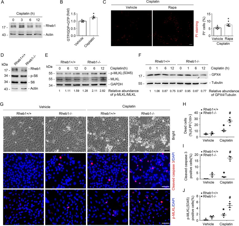
Fig. 5. Ablation of Rheb1 in primary cultured tubular cells exacerbates cisplatin-induced cell death.
a Western blot assay showing the induction of Rheb1 in primary cultured tubular cells incubated with 25 mg/ml cisplatin for 3, 6, and 12 h. b GTP loading assay showing the induction of GTP-Rheb1 after cisplatin treatment at 3 h. *P < 0.05 vs. Vehicle control, n = 4. c PI staining and quantitative analysis of dead cells after cisplatin treatment. Data is presented as the percentage of PI-staining positive cells. *P < 0.05 vs. vehicle control cells, n = 5. Scale bar = 20 µm. d Western blot assay showing the reduction of Rheb1 and p-S6 abundance in primary cultured tubular cells with Rheb1 ablation. The primary cultured tubular cells from Rheb1fl/fl mice were infected with adenovirus carrying GFP or Cre recombinase gene for 48 h, respectively. e, f Western blot assay of p-MLKL (e) and GPX4 (f) in primary cultured tubular cells after cisplatin administration. g Representative micrographs showing the cell detachment, anti-cleaved caspase 3 and anti-p-MLKL staining as indicated. Scale bar = 20 µm. h–j Quantitative analyses of dead cells, cleaved caspase 3-staining positive and p-MLKL-staining positive cells among groups as indicated. Data is presented as the percentage of the staining positive cells. *P < 0.05 vs. vehicle control, #P < 0.05 vs. Ad-GFP-infected cells treated with cisplatin, n = 3–4.

